Interest in characterising soil communities is booming, fuelled by the growing recognition that soil biota govern processes of carbon (C) and nitrogen (N) cycling – processes that underpin the delivery of soil-based ecosystem services such as climate mitigation and sustainable food production. Soils capture carbon, which can exacerbate climate change when released to the atmosphere, and they provide nitrogen and other nutrients for growing crops and feeding livestock – when these nutrients are lost from soils, they can pollute ground and surface water and cause a loss of biodiversity. Because soil microbes decompose organic matter, thereby releasing N for plant growth, and respiring C, they determine the balance between the release and retention of C and N in soils.
In my work, I have a particular interest in the role of soil fungi and bacteria in these processes. Moreover, I want to find out how land use change and climate change affect the relative abundance of fungi and bacteria, and the chain of soil fauna that feed on them (the fungal and the bacterial energy channel, respectively), and how these changes in turn affect processes of C and N cycling. For example, some of my recent work shows that fungal-dominated microbial communities of extensively managed grassland retain N better and have lower N leaching losses, about which you can read more in this old blog post. Also, I have shown that fungal-based soil food webs and the processes of C and N cycling that they carry out are less affected by drought, which is expected to increase with climate change, than bacterial-based soil food webs.
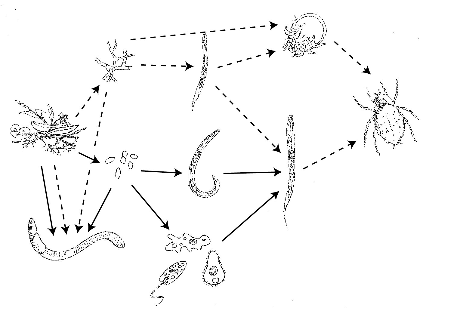
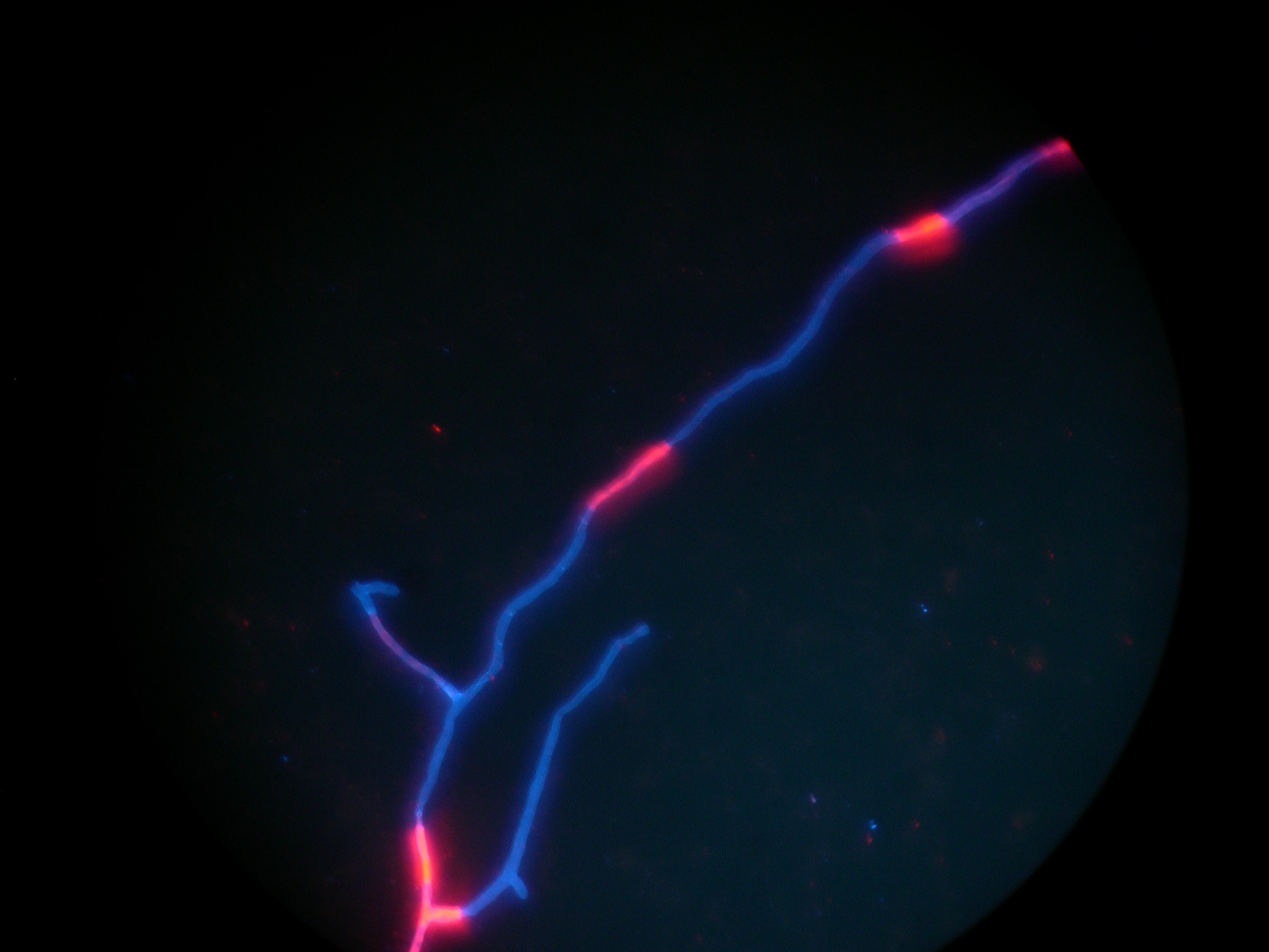
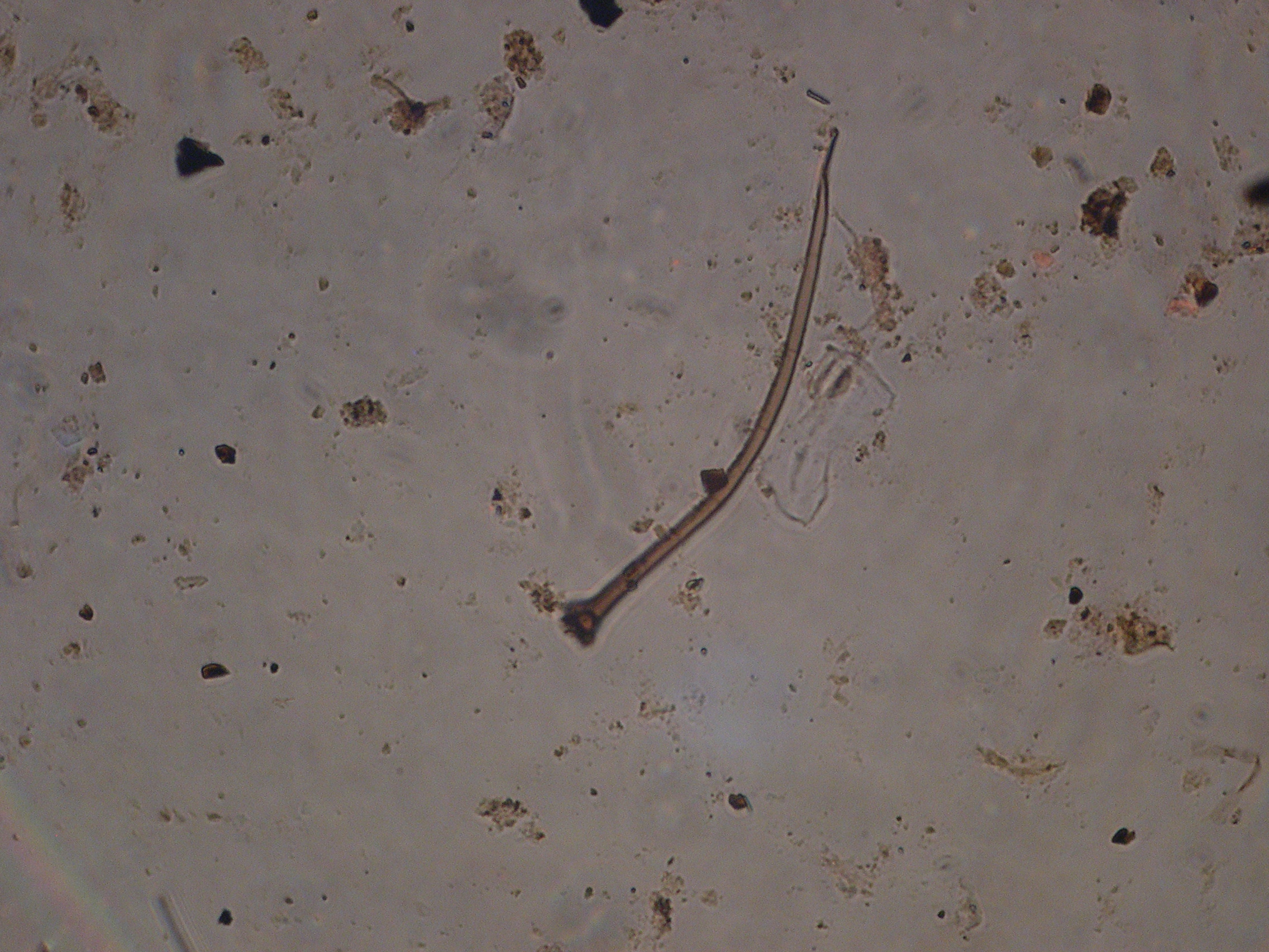
To do this type of work, obviously, you have to measure the composition of soil microbial communities, or even of entire soil food webs. This is not an easy task, as most of these organisms are not, or barely, visible for the naked eye. For decades, direct microscopy was the only possibility to quantify and characterise the composition of soil microbial and soil faunal communities. For microbial communities, this involves transferring a soil suspension onto a microscopic slide, staining the fungi and bacteria, and then counting their hyphae or cells using a microscope. I used this method during my PhD and spent weeks, if not months, looking through a microscope. Although still frequently used, in recent years, direct microscopy has been increasingly replaced by the measurement of phospholipid fatty acids (PLFAs), a component of the cell membranes of fungi and bacteria. Because different microbes have different PLFAs in their cell membranes, the PLFA composition of a soil sample can be used as a ‘fingerprint’ of the soil microbial community. In other words, it doesn’t only tell you about the relative abundance of fungi and bacteria, but also about the composition of the bacterial community.
Although PLFA analysis is relatively new (it was first used in the 1980s on aquatic samples), increasingly, molecular methods are used to characterise microbial communities. These methods have opened up a whole new realm of ways to characterise soil microbial and faunal communities, from functional properties, to number of gene copies of a certain group, to the entire genome in a soil sample. Although these analyses are expensive, they are becoming cheaper, and they allow for the analysis of large numbers of samples. For example, in a recent paper in PNAS, Wu et al. analysed 17,516 18S rRNA gene sequences to describe faunal communities in soil samples from across the world. In another study, Griffiths et al. analysed over 1000 soil samples using T-RFLP to assess bacterial biogeography in the UK.
So, both PLFA analysis and molecular methods have obvious benefits over microscopy. However, they also have one major drawback: you can’t see what is going on. Both methods consist of a lengthy procedure in which you have to extract PLFAs, DNA, or RNA from soil samples. This involves a large number of steps – weighing, adding reagents, vortexing, centrifuging, pipetting – in which you can’t see whether you are doing it right. Also, these extractions often involve nasty substances, such as hexane, chloroform, or ethidium bromide. Moreover, at the end of the analysis, you end up with a graph with peaks that represent concentrations, or even just a file with numbers.
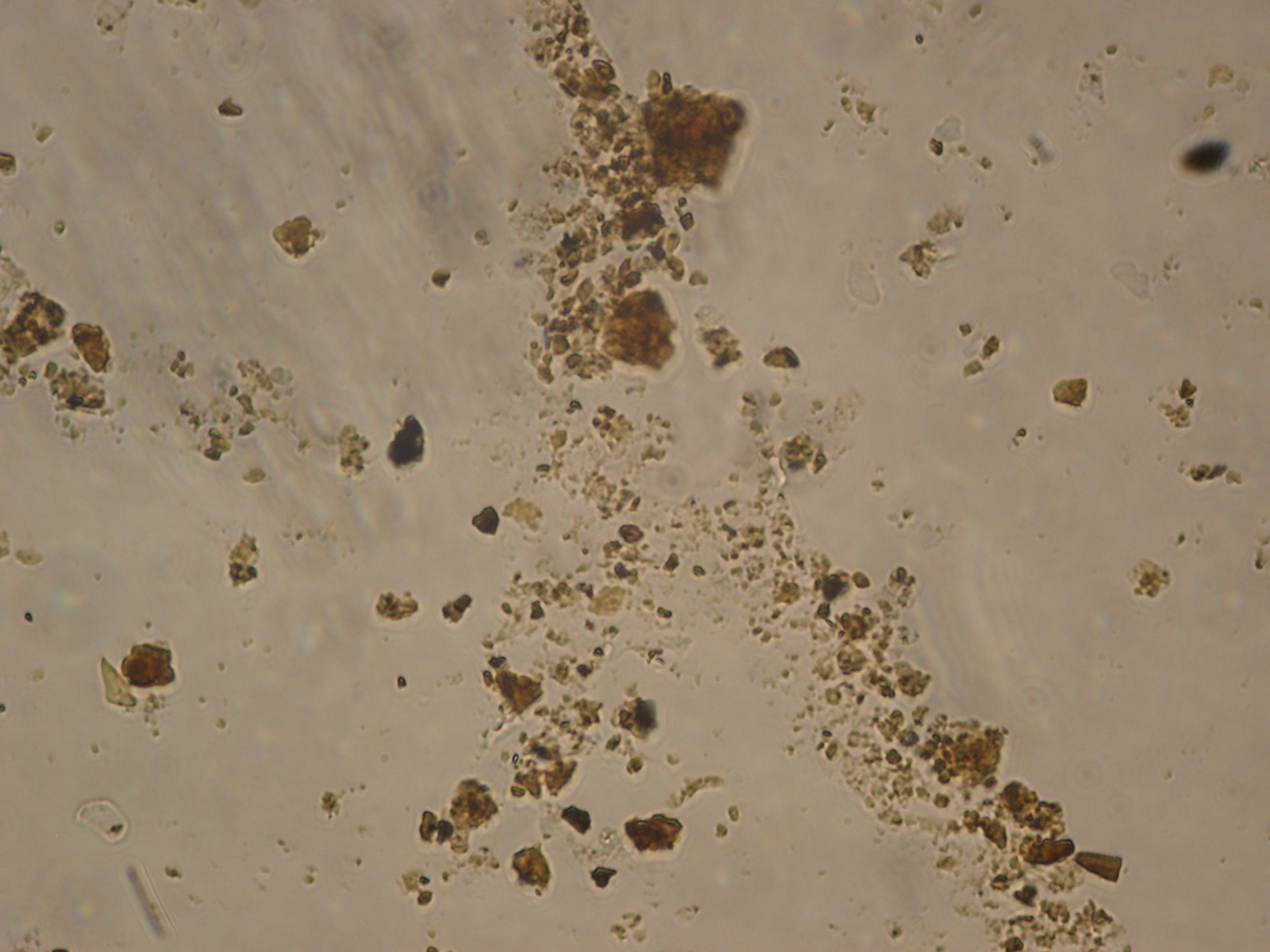
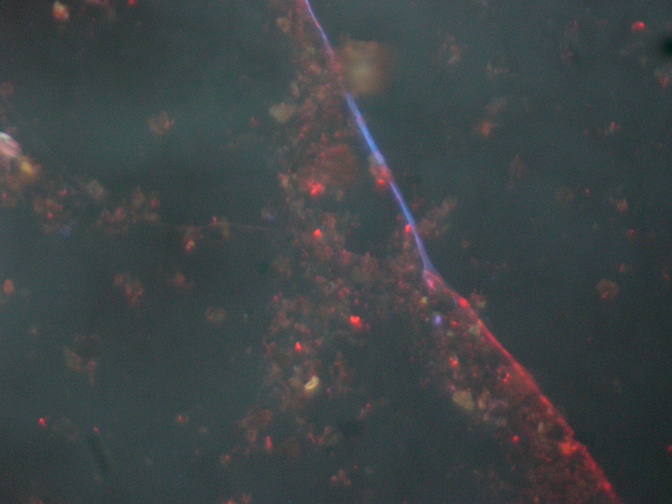
In contrast, traditional, microscopic methods allow you to see the actual organisms that you are studying, which are frequently stunningly beautiful. During the many hours I spent looking through a microscope and counting fungal hyphae, I saw hyphae attacking a nematode (a microscopic small worm), a tardigrade (see this post for a picture of the cutest soil animal alive), hyphae with exploding spore capsules, hyphae entangling soil particles, penetrating roots, and hyphae with millions of bacterial cells all over their surface. A microscope allows you to see how microbes interact with their environment, something that the most advanced molecular method will never let you experience. Right now, a whole generation of soil ecologists is being trained that has never seen a fungal hyphae or a bacterial cell. Although they learn about interactions between microbes and each other, and between microbes and their environment, seeing these phenomena with your own eyes makes a world of difference. If only more people would be aware of the beauty and diversity of life in soil, I think we would treat our soils a lot better.
These days, microscopy, and even PLFA analysis, are perceived to be out-dated, and there is a real tendency to use these high-throughput molecular techniques wherever possible. However, in my opinion, if you can answer your research question using a simple method, why use a complicated, and expensive, method? If you want to know how bacterial cell size is affected by land use or climate change, simple microscopy will do, and if you want to know about broad shifts in soil microbial community composition, PLFA analysis is sufficient. For more mechanistic questions, or if you want to identify the exact species in your sample, it might be a good idea to use molecular techniques.
So, the morale of this post? Every method for studying soil microbial communities has its benefits and drawbacks. Although microscopy and PLFA analysis might seem out-dated, they are still valid techniques. Moreover, microscopy shows you the actual organisms and their interaction with their environment – something that is of crucial importance to make people aware of the diversity of life that soils harbour.

Leave a reply to ibartomeus Cancel reply